Sample Preparation of Plant and Animal Tissue in Remote Locations: Tools and Methods
By Tim Hopkins, PhD
Molecular biology methods are increasingly being used to monitor agricultural products directly on location where the crops and animals are commercially produced. By doing so, problems such as pathogens and disease, chemical contamination, and effects of environmental stress receive prompt solutions. The sooner the farmer detects and applies needed steps to rectify a problem, the better the outcome.
The traditional workflow for molecular biology testing in the field can be broken down to several steps:
- A suitable sample is taken from the plant or animal.
- The tissue or its intracellular components such as DNA, RNA, and proteins are stabilized for delivery to the centralized laboratory. Stabilization is often done by freezing or icing-down the sample. This part of the protocol is time-sensitive and susceptible to unanticipated handling mishaps.
- The homogenate or tissue extract is then analyzed in the centralized laboratory using specialized instrumentation such as GC-, LC-, ICP-MS and qPCR, and the results with their conclusions are relayed back to the farmer.
An active area of development is the availability of improved hand-held tissue extraction tools and accompanying reagent kits designed for use in field locations. With these assets, tissue samples can be harvested, stabilized on site, and delivered by conventional courier to the distant analytical laboratory without refrigeration or cooling requirements. The benefits are clear. The analytical laboratory receives consistently high quality tissue extracts and the farmer receives needed information with a minimum of delay, thus providing significant savings in operating costs.
Once the tissue is harvested, molecular biology protocols often call for on site dispersion or disruption of the harvested tissue. This is usually done by dispersing the tissue sample using a tissue press or, if the goal is analysis of nucleic acids, grinding the tissue in a microvial containing extraction solution and small spherical beads. The cell extraction may then be stabilized with added chemicals or transferred to a special stabilizing filter paper.
Tissue Harvesting Tools
Tissue harvesting begins with removing a tissue sample from a plant or animal using a knife, scissors, or tissue hole punch. For most tools used, an adequate sample size is generally less than a gram of material. The tissue should be handled using sterile techniques, which includes using surgical rubber gloves and clean tools. Between use decontamination of the tools can be done using either 70% alcohol, household bleach (1 part in 10 parts water), or heating in boiling water.
Tissue Hole Punches
Hole punches are especially convenient because the sample size is reproducible. Unfortunately, punched samples are limited to thin, flat tissues such as plant leaves, animal ears and fish fins.
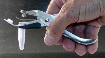
Biospec Products (Bartlesville, OK) makes a leaf tissue punch that goes a step further. Their punch holds a standard 1.6 or 2 ml microvial in a fixed position to receive a 3/8 inch diameter disc of tissue without further manipulation.
If homogenization or cell disruption of the tissue sample is the end goal, the tissue sample can often be reduced in size by chopping it into pieces less than 1 mm in cross section with scissors or single-edge razor blade. Finely chopped tissue homogenizes faster.
Tissue Presses
Some tissue samples can be reduced even further in size by dispersion in a screw- or lever-tissue press.
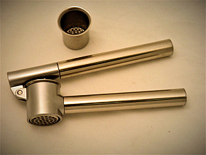
The LeverTissuePress™ (BioSpec Products) macerates or mechanically disaggregates 5 to 50 grams of fresh, soft plant or animal tissue by forcing the tissue through an array of tiny holes. Acceptable tissues to use are muscle, brain, liver, plant fruits, and some plant roots and tubers like potatoes and carrots.
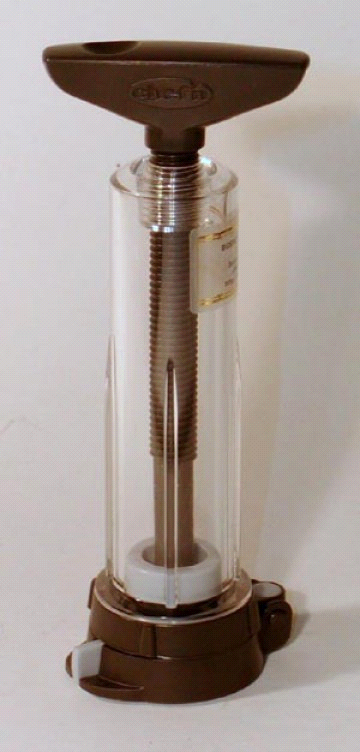
The ScrewTissuePress™ (BioSpec Products) dispersess 0.5 to 10 g of tissue by forcing soft tissue through screens having 0.250 micron or 2 mm diameter holes. No buffer or dispersion media is required for this process. The dispersed tissue exits the ScrewTissuePress having a consistency resembling liver paté and still remains mostly viable, intact cell material.
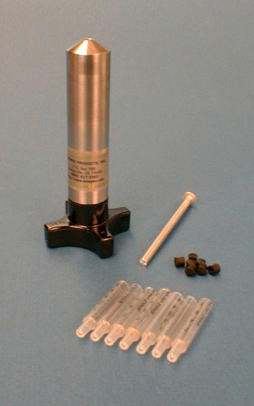
The MicroMincer™ (BioSpec Products) is a smaller, hand-operated screw press which macerates 0.3 grams or less of soft plant or animal tissue (e.g. muscle, liver, brain, epicotyl and pulpy leaf tissue). The forces it can develop to push tissue through a 250 micron screen are much higher than the ScrewTissuePress. Like the ScrewTissuePress, the tissue emerges from the MicroMincer having the consistency of liver paté.
Compared to the ScrewTissuePress, the MicroMincer can process tougher, more fibrous tissues, such as leaf, stem, and root, and animal sample having high connective tissue content. The pressed sample is extruded out of the MincoMincer's tip as a clarified intracellular extract while the fibrous pulp and cell wall material remains behind, embedded on the screen.
The MicroMincer utilizes disposable polypropylene liners. Thus, multiple samples can be processed without a need to reclean the device or risk contamination between samples.
Leaf Crushers
A new approach to taking a tissue sample in the field is crushing a portion of the tissue using high compressive forces. Currently, the method applies to flat, thin tissue such as leaf tissue and fish fins. During the crushing process, released intracellular cell contents are captured and stabilized by adsorption on cellulose filter paper. Later, at the analytical laboratory, the captured cell contents are released from the filter paper in a partially purified form.
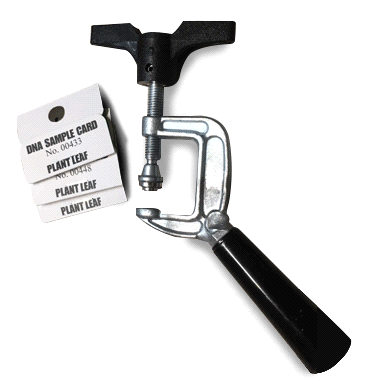
The MiniLeafCrusher™ (BioSpec Products) is a modified C-clamp designed to crush open tissue cells of the sample and release the nucleic acids of the plant and possible intracellular pathogens. Used in combination with a unique DNA Sample Card™ (BioSpec Products), the leaf is inserted inside the folded card, which is magnetically docked into the clamp where a 1/4 inch diameter area of the leaf is crushed at thousands of pounds per square inch. The released cell extract is immediately captured and stabilized on special cellulose filter paper within the DNA Sample Card. After briefly drying in air, the nucleic acids adsorbed on the filter paper are stable for months without refrigeration. The nucleic acids can be eluted from the filter paper in minutes and amplified using PCR techniques for sequencing.
The DNA Sample Card alone can also be used to stabilize and transport a drop of blood, sputum, urine or, as discussed below, homogenized tissue samples disrupted by grinding.
Tissue Cell Disruption Tools
Tissue samples can be mechanically disrupted by grinding the sample in microvials containing extraction media and grinding beads. The beads are made of glass, ceramic, or steel and vary in size from 0.1 mm to 3.2 mm in diameter. The homogenate can be stored on the BioSpec DNA Sample Card or stabilized with special preservative solutions that accompany commercial nucleic acid isolation kits.
Pestle and Tube
Disruption of tissue cells by grinding is a traditional method. Pestle and microtube grinders come in various sizes and are available from numerous suppliers. They function by forcing tissue to repeatedly pass through the narrow clearance between the surface of the pestle and the inside surface of the tube. For small tissue sample sizes, most tube and pestle grinders are 1.5 ml conical polypropylene microtubes paired with matching pestle that are customarily operated by hand-rotating the pestle shaft.
A recent improved version of the smooth-sided polypropylene pestle is the SpiralPestle™ (BioSpec Products). Combined with the a standard 1.5 ml conical microtube, it homogenizes small samples of soft plant and animal tissue and is especially effective when a few 1 mm diameter glass beads are included during use. The 'threads' in the turning Spiral Pestle act like a tiny screw pump. It drives 10 to 100 mg of soft plant or animal tissue suspended in 200 to 500 µl of extraction solution to the bottom of the microvial where it is macerated and mechanically dispersed.
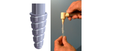
When rotation of the SpiralPestle is powered with a battery-driven BioVortexer™ mixing motor (BioSpec Products), homogenization can be completed in seconds.
Beadbeater Cell Disrupter
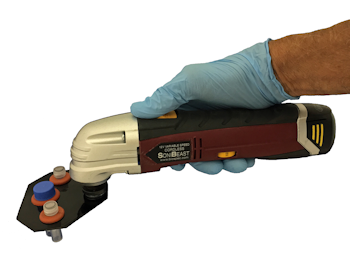
Beadbeating (also called shaking bead milling) has become the preferred way to mechanically disrupt the cells of all types of tissue. The portable SoniBeast-Jr™ is a hand-held version of BioSpec Products' series of laboratory beadbeaters. Powered by rechargeable batteries, the high energy beadbeater cell disruptor holds either two 2 ml microvials or one 7 ml vial. Filled with ceramic beads and extraction media, the SoniBeast-Jr can completely disrupt 10 to 400 mg of plant and animal tissue in less than a minute.
What's Coming Next for On-Site Tissue Sampling?
Analytical services which must currently be provided by centralized laboratories may no longer be needed. Instead, with easy-to-use reagent kits and compact, hand-held sensors, the farmer will have access to instant analytical information. These specialized analytical tools and methods will come in forms ranging from a simple dip-stick that displays a color when placed in a tissue extract to a hand-held electrochemical monitor that wirelessly connects to a cell phone app.
Although specific kits and devices are not yet available in the agriculture market, fundamental technologies are in place to make this a reality. For example, consider the familiar compact household analytical devices used to monitor pH in a spa, glucose concentration in a blood sample, or a hormone in a pregnancy test.
This editorial was written by BioSpec Products and published in collaboration with LabX.
