Microscopy Frontiers: Pioneering Innovations Transforming Scientific Imaging
Introduction
The world of microscopy is undergoing a remarkable transformation across biology, materials science, and beyond, democratizing access to powerful imaging tools and pushing the boundaries of discovery. Here we explore several fresh developments that are reshaping how we see the microscopic world.
New Microscopy Method Enables Protein Imaging with Conventional Light Microscopes
Researchers at the University Medical Center Göttingen have developed One-step Nanoscale Expansion (ONE) Microscopy, a groundbreaking technique that allows imaging of 3D protein structures using conventional light microscopes. This method expands biological samples up to 15 times their original size by embedding them in a water-absorbing gel, making nanoscale details visible beyond the optical resolution limits of traditional fluorescence microscopy.
By combining ONE microscopy with artificial intelligence, scientists reconstructed the 3D shapes of individual proteins, such as the Y-shaped antibody structure, previously achievable only with costly technologies like cryo-electron microscopy. The method has already demonstrated potential in detecting toxic protein aggregates, such as alpha-synuclein linked to Parkinson's disease, from patient cerebrospinal fluid samples. This innovation offers affordable, high-resolution imaging and holds promise for early diagnosis of neurodegenerative diseases and other protein misfolding disorders.
A New Technique Brings High-Resolution Imaging to Conventional Microscopes
MIT researchers have developed an accessible and cost-effective method to achieve nanoscale resolution using standard light microscopes. This innovative expansion microscopy technique, described in Nature Methods, allows biological tissue to swell 20-fold in a single step, enabling detailed imaging of structures as small as 20 nanometers without requiring expensive super-resolution microscopes.
By embedding tissue in a specially optimized polymer gel and expanding it with water, scientists can visualize cellular components like organelles, protein clusters, and nuclear pore complexes. This single-step process simplifies earlier multi-expansion methods while maintaining high resolution. The approach uses common lab equipment and affordable chemicals, making high-resolution imaging accessible to more biology labs.
Applications include studying brain synapses, cancer cell structures, and cell-surface glycans, providing unprecedented insights into nanoscale biological processes. This technique democratizes high-resolution imaging, empowering labs worldwide to explore cellular mechanisms in greater detail than ever before.
New 3D Microscopy Method Enables RNA Analysis in Whole Brains
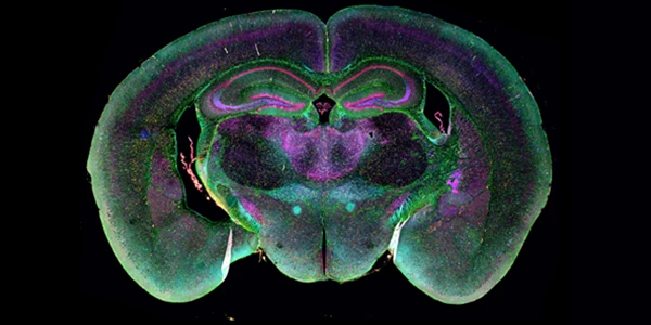
Researchers at Karolinska Institute and Karolinska University Hospital have developed TRISCO, a novel microscopy technique allowing 3D RNA imaging at cellular resolution in whole, intact mouse brains. Published in Science, TRISCO eliminates the need to slice tissue, addressing a longstanding challenge in RNA analysis by preserving spatial context.
The study demonstrated simultaneous visualization of three RNA molecules, with plans to expand to multiplex analysis of around 100 RNA molecules, offering deeper insights into brain function and disease mechanisms. TRISCO is also adaptable to larger brains, such as guinea pigs, and other tissues, including kidney and lung, broadening its applications.
This breakthrough has the potential to revolutionize neuroscience and lead to novel treatments for brain diseases. The collaborative effort underscores the importance of bridging basic research with clinical expertise to advance biomedical innovation.
Scientists Visualize Bacterial Protein Synthesis Initiation for the First Time
An international research team, including scientists from the University of Michigan, has unveiled how ribosomes in bacteria recruit and interact with mRNA during transcription. Using advanced cryo-electron microscopy (cryo-EM) and other techniques, the researchers revealed that RNA polymerase (RNAP), the enzyme transcribing mRNA, employs dual "anchors" to secure the ribosome, ensuring efficient protein synthesis.
This study offers a first glimpse of how RNAP and ribosomes coordinate in bacteria, where transcription and translation occur simultaneously. The findings provide a framework for understanding how newly transcribed mRNAs are delivered to ribosomes for translation, potentially enabling the development of novel antibiotics targeting these critical pathways.
By identifying alternative mechanisms of mRNA delivery and ribosome coupling, the research paves the way for targeting bacterial protein synthesis with precision, overcoming resistance seen in existing antibiotics. The work, combining cryo-EM, crosslinking mass spectrometry, and fluorescence microscopy, marks a breakthrough in visualizing the earliest stages of gene expression.
Microscopy Advances Pave the Way for Dynamic Chemical Reaction Imaging
A groundbreaking collaboration led by the University of Illinois Chicago (UIC)and other institutions aims to transform our understanding of chemical reactions at the atomic level. The new Center for Multimodal Observations for Single Atom Imaging of Chemistry (MOSAIC) is funded by a $1.8 million National Science Foundation (NSF) grant, with $270,000 allocated to UIC. The initiative seeks to develop advanced electron microscopy techniques to observe chemical reactions in real time.
Key to this innovation is the use of graphene "liquid cells," which shield samples from the damaging effects of electron beams, enabling researchers to capture undisturbed, dynamic reactions. UIC's state-of-the-art electron microscopes, including a first-of-its-kind magnetic-field-free microscope set to debut in 2025, will play a critical role in this effort.
Initially, the research will focus on single-atom catalysts and small molecular clusters, with aspirations to analyze complex molecules and critical processes in chemistry, biology, and materials science. The project builds on earlier advances in graphene-based imaging and positions UIC as a leader in electron microscopy, fostering collaborations across the wider scientific community.
JEOL-IDES Licenses Advanced UTEM Technology for Breakthrough Applications
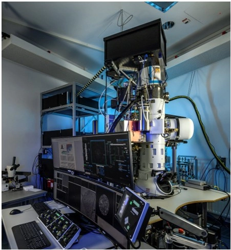 JEOL-IDES has licensed ultrafast transmission electron microscopy (UTEM) technology from
the Max Planck Institute for Multidisciplinary Sciences and the University of
Göttingen. Developed by Professor Claus Ropers' team, this innovation
integrates laser-driven electron emitters into TEM systems, enabling
unprecedented precision and speed for imaging at the atomic scale.
This advancement enhances resolution and allows real-time observation of ultrafast processes, unlocking new possibilities in materials science, biology, and nanotechnology. The collaboration bridges cutting-edge research and industrial application, ensuring the technology's accessibility and continual refinement.
Facilitated by Max Planck Innovation and MBM ScienceBridge GmbH, the partnership exemplifies the power of academic-industry cooperation to deliver transformative tools for scientific and industrial innovation worldwide.
Bruker Launches Dimension Nexus™ AFM for Cost-Effective High-Performance Imaging
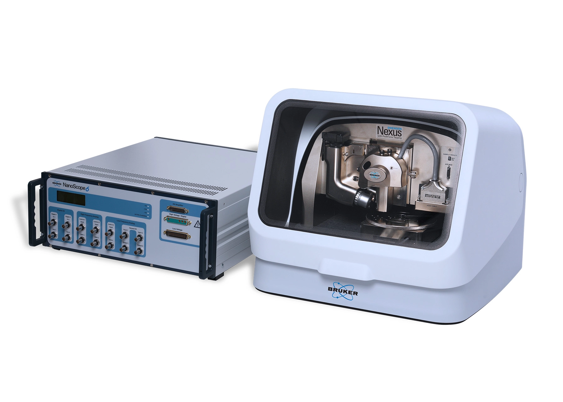 Bruker has introduced the Dimension Nexus™ atomic force microscope (AFM) at the 2024 MRS Fall Meeting. This latest addition to the Dimension® AFM product line
features the state-of-the-art NanoScope® 6 controller, offering enhanced
performance, upgradability, and ease of use. Designed for both routine and
custom applications, Dimension Nexus provides advanced imaging capabilities
with low drift, low noise, and over 50 AFM modes, including PeakForce Tapping®
and AFM-nDMA for viscoelastic measurements.
The system’s open architecture, compact design, and programmable stage enable high-throughput, multi-site analysis, making it an ideal solution for both growing labs and multi-user facilities. Dimension Nexus sets a new standard for accessible, versatile AFM technology, combining high-quality data acquisition with cost-effectiveness for evolving research and industrial needs.
