Magnetic Resonance Imaging of the Brain: Unprecedented Power and Resolution
Magnetic Resonance Imaging (MRI) has evolved as a highly versatile technique for brain and organ imaging. As a medical relative to nuclear magnetic resonance (NMR) spectroscopy, increases in resolution have come with the advent of larger magnets and innovations in performance. The latest MRI systems have now surpassed the 10 Tesla mark -- making clear the power of the technology for cutting-edge brain research and advanced diagnostics.
Nuclear Magnetic Resonance Spectroscopy
The underlying physical method, NMR spectroscopy, is a spectroscopic technique typically applied towards molecular-scale structural studies. Pioneered in the late 1940’s by investigators at Harvard and Stanford universities, NMR garnered the 1952 Nobel Prize in Physics for its discoverers, Edward Mills Purcell and Felix Bloch.
Principle of NMR spectroscopy
In basic terms, NMR can detect and measure local magnetic fields around atomic nuclei. Organic ions (hydrogen or carbon) in a sample are excited by exposure to a magnetic field causing them to align in accordance with their charge and the direction of the field. The NMR source emits a short radio wave burst causing the atoms to oscillate. The resulting nuclear magnetic resonance is then detected by sensitive radio receivers. Intramolecular magnetic fields around the atoms of a molecule influence the resonance frequency of a given atom, thereby providing details on the local structure of each nucleus.
Proton NMR
The most common types of NMR are proton or 1H and carbon-13 NMR – although the technique is applicable to any sample containing nuclei possessing spin. Although most organic molecules contain a suitable number of NMR active hydrogen atoms, analysis can be challenged by the large abundance of protons present in organic solvents. One way around this is by using deuterated solvents, where >99% of 1H atoms are replaced with 2H deuterium in solution. Another method involves biosynthesis and purification of the biomolecule of interest sourced from (micro) organisms propagated in deuterated growth media.
NMR analysis
Differing electronic environments in a molecule of interest are resolved by the applied magnetic field and measurement of the resulting magnetic resonance. “Local” effects shield protons - to differing degrees - from the large external applied field. These chemical shifts in resonance energy are very small, on the order of parts per million, and can be used to determine molecular structure through a process called assigning the structure.
NMR instrumentation
NMR Spectrometers are highly sensitive and complex devices – and therefore expensive in terms of instrument costs, facilities design, and maintenance. Modern NMR instruments posses superconducting magnets which are large, expensive, and require liquid-helium as a coolant. Resolution depends on the strength and performance of the magnet, so this aspect of the instrument cannot be compromised.
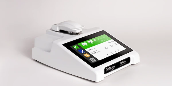
There are much smaller bench top NMR spectrometers for research applications, which can offer a valuable alternative to other analytical techniques and enhance the NMR workflow in an analytical lab. These systems are often well-suited for chemical analysis, structural studies, and materials testing. They also offer viable solutions to issues of larger machines such as: lack of availability, time requirements of analysis, costs of outsourcing, lack of in-house expertise, and other limitations.
Nuclear Magnetic Resonance Imaging
Generally speaking, the stronger the magnet, the greater the fraction of protons that align – and the greater the resolution. There is a lag however, between the jump in field strength of an NMR platform and implementation for magnetic resonance imaging.
Remember, MRI is the medical application of NMR and, although sample conditions and consistency can be carefully controlled in the lab and sample tube, the same cannot be said for imaging intact tissue in the body. Early concerns of MRI included whether the radio waves – and resulting resonance signals – could effectively penetrate tissue of varying density and complexity, such as brain structures. This, along with the concerns of limited resolution, formed the driving force for larger more powerful MRI devices for human imaging.
New dimensions of MRI in research and medicine
In the 1980’s, 1.5 Tesla (T) scanners came into clinical use. In 2003, 3-T MRI. Now we are amidst a breakthrough to 7-T scanners and the myriad of biological events which can now be imaged in the brain.
Of course, there are practical considerations that need to be addressed. For instance, involuntary movements such as breathing and patient heart beat – even movement of body parts in remote areas and connected through the spine to the base of the skull – can affect imaging at this new enhanced resolution. Other issues including “hot spots’ resulting from increased field strengths, and corresponding instrument design features, must be addressed to prevent tissue damage and imaging distortions.
Nonetheless, this new level of MRI power offers promise for exploring the biological basis of memory and speech, disease mechanisms such as Multiple Sclerosis and Alzheimer’s, and for increased accuracy and precision of deep brain stimulation, microsurgery, and other therapeutic applications.
Is there an upper limit to MRI magnetic field strength and imaging resolution? Heat and noise generation may continue to be complicating factors – although work in animals has pushed the envelope to 21.1-T with seemingly no ill-effects. Regardless of the complications, however, it’s clear that the innovation of NMR instrumentation will continue to lead the way towards new thresholds of MRI power and resolution.
