Cell Imaging and Screening Applications Enabled by Advanced Microplate Technologies
As reviewed previously, modern microplate instrumentation has turned single purpose instruments into multiplexing platforms, which in essence has brought many distinct testing technologies together under one lid
In this article we explore novel applications and new products which are enabling breakthrough research in microscopy, stem cell biology, high-content/high-throughput screening, and other areas.
Microplate Cell Imaging
The concept of microplate-based analysis arose from gains in detection sensitivity, giving rise to higher throughput multiplexing capabilities with lower sample volume requirements. Light scattering, UV-Vis absorbance, and other spectroscopic techniques were the norm for measurement.
With the advent of advanced microscope technologies came the ability to glean physiological information from cells under native-like conditions. Modern technologies evolved to merge microplate-based detection and cell imaging microscopy into a central concept – creating enabling solutions for research, drug discovery, and other important applications.
Live Cell Imaging
Although challenging, live-cell imaging is very useful for many aspects of drug development such as cell motility or movement and kinetic studies of compound mode of action. Research applications of live-cell imaging include cell-division, apoptosis, and toxicity studies, among others.
Automated Microscopy
Breakthroughs in automated microscopy and image analysis have led to the establishment of imaging platforms as screening tools.
High Content Analysis
Dramatic improvements in imaging resolution and data analysis have translated to increases in the quality and quantity of cellular information – the basis of high-content analysis (HCA).
High-Content Screening
High-content screening (HCS) arose as a platform to capture enhanced physiological relevance of cellular activity as opposed to biochemical information and traditional routes of analysis. As a result, HCS has become the ideal and preferred method for accelerating modern drug screening campaigns.
High Content Imaging Systems
Although most high content imaging studies are performed using fixed cells, emerging technologies are focusing on live cells with features designed to address the challenges associated with cell culture and cell viability demands. Many automated microscopy vendors offer incubation, liquid handling, onboard pipetting, and other features essential to propagation and manipulation of live cells – in concert with automated microscopy and HCA/HCS techniques.
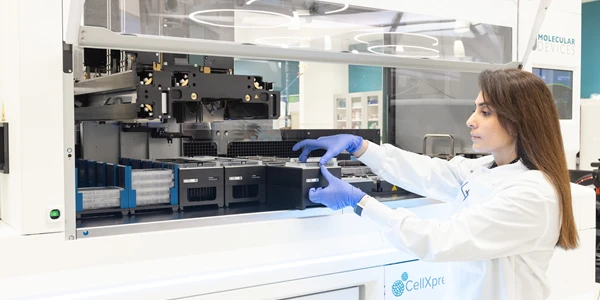
Molecular Devices offers a range of cutting-edge instruments designed for high-content or high-throughput lab applications.
- The ImageXpress Micro Confocal high-content imaging system is capable of capturing high-quality information and 3D reconstruction of cells – with throughput of over 1 million wells per week.
- The ImageXpress Micro 4 is a wide-field imager for high-throughput acquisition of whole organisms and cellular or intracellular events. Options include the availability of confocal, brightfield, phase contrast, liquid handling, environmental control – all highly configurable and flexible.
- The ImageXpress Nano is a high-content imaging system for everyday biological needs in the lab. A flexible solution for fluorescence-based cellular analysis.
The above platforms all use the MetaXpress high-content image acquisition and analysis software, which allows 2D and 3D imaging, time lapse analysis, programmable methods, and use of hundreds of preprogramed routine assays. 4D imaging combines confocal with time-lapse microscopy to enable high-resolution live-cell analysis.
- The ImageXpress Pico combines high-resolution imaging and analysis with a straight forward workflow. The CellReportXpress software features a portfolio of preconfigured protocols. The automated setup features accelerate the rates from start to result. The system is an affordable entry point for fluorescence imaging and digital microscopy.
Advanced applications for these platforms may include: Organ-on-a-chip assays, stem cell growth and differentiation, toxicity screening and proliferation assays, and many more techniques.
BioTek offers a host of automated cell imagers for varied applications.
- The Lionheart automated microscopes are designed to capture images, z-stacks, montages, and time-lapse sequence. Brightfield, color brightfield, phase contrast, and fluorescence channels support a range of workflows.
- Live cell imaging is enabled with the Lionheart FX, with optional 40° CO2/O2 and humidity control.
- The Gen5 software supports Augmented Microscopy – automated image capture, processing, and analysis to produce publication-ready images and data.
- The BioSpa Automated Live Cell Imaging System integrates the BioTek Cytation Cell Imaging Multi-Mode Reader with the BioSpa Automated Incubator. Liquid handling can be added to produce a complete system, from sample prep to image analysis.
- The Cytation 5 and Cytation 1 Cell Imaging Multi-Mode Readers combined automated microscopy with traditional microplate reader platform – with the flexibility of modular upgrades. These include: fluorescence intensity, UV-Vis absorbance, luminescence, fluorescence polarization, and time-resolved fluorescence and laser-based alpha.
Live cell imaging, toxicity and apoptosis, cell proliferation, stem cell differentiation and 3D cell imaging are all possible.
Emerging automated microscopy and microplate technologies are focusing on novel imaging approaches.
For instance, Event Driven Automated Microscopy diverges from traditional approaches in that sample variations drive the measurement parameters. The Z-stack, imaging channel(s), position(s), view settings and more are defined by the nature and dynamics of the sample itself. The method avoids bias and allows larger scale studies without loss of search space or critical information. Cancer research is an application where time-lapse imaging of cell mitosis and cell proliferation may give rise to important phenotypes and migrations, enabled by this advanced technique.
View imaging system listings at LabX.com
